






Osteomas are benign bony growths that can develop on the skull, often around the forehead or ear region. Removal of osteomas may be considered in cases where they cause physical discomfort, limitations with headwear, glasses, or helmets, or are visibly prominent. Once an osteoma has been confirmed by a specialist, patient suitability for removal can be assessed.
All osteoma removals at the CREO Clinic are performed in CQC-registered facilities by our Medical Director, Dr Omar Tillo, who is a GMC-registered Plastic Surgeon, a Fellow of the Royal College of Surgeons (FRCS), and a member of the British Association of Aesthetic Plastic Surgeons (BAAPS). Dr Tillo regularly teaches various surgical techniques to other BAAPS members and has led the creation of UK guidelines for key body contouring techniques.
Surgery at a Glance
Anesthesia
Local or generalSurgery Time
Between 30 min – 2 hoursHospital Time
Day caseOff-Work
1 – 2 daysWhy Patients Consider Osteoma Removal
Patients may seek specialist assessment for an osteoma in cases of physical discomfort or visibly prominent lumps. Assessment can also help clarify the nature of the lump and determine whether further investigation, monitoring, or osteoma removal may be appropriate.
Visible Forehead or Skull Prominence
An osteoma typically presents as a hard, immovable lump beneath the skin, often on the frontal bone, scalp, or around the ears. In some cases, an osteoma can be concealed or barely noticeable, whereas some osteomas can be large enough to create significant irregularity in the contour of the skin around the head.
Any irregularity in contour caused by the osteoma can offset symmetry and negatively impact the facial profile; an osteoma removal may help improve any cranial contour changes.
Discomfort or Pressure Symptoms
Depending on their position or severity, osteomas can cause discomfort in multiple ways. Glasses frames, helmets, hats, or any direct contact pressure can all create localised tenderness at the osteoma site. For many patients, these physical symptoms are a primary reason for seeking assessment and osteoma removal.
Diagnostic Certainty and Peace of Mind
Another important concern around osteomas is whether the lump is benign or requires further assessment. Specialist assessment by an experienced surgeon, sometimes supported by CT or X-ray imaging, can help establish diagnostic clarity and guide appropriate next steps before any surgical plan is considered.
Understanding Osteomas
Understanding exactly what an osteoma is and how it differs from other types of unexplained lumps can help patients understand the likely diagnosis, consider whether osteoma removal may be appropriate, and make informed decisions about their treatment. Below, we take a look at osteomas and their associated concerns in more depth.
What Is an Osteoma?
An osteoma is a usually benign bony growth that develops when new bone tissue forms on an existing bone. These lumps are slow-growing and immovable, often appearing in areas around the skull, on the forehead, and on the jaw. Their exact cause remains unclear, although it is believed that genetic factors and developmental anomalies may be associated with osteoma growth.
There are three main types of osteoma: the peripheral type, which sits on the surface of the bone; the central type, which is found within the bone; and the extra-skeletal type, a very rare type that can be found in the soft tissue.
Are Osteomas Dangerous?
In general, most osteomas are benign and do not represent an immediate health concern; however, they can be a source of irritation and physical discomfort. Any new, changing, painful, or unexplained lump should be assessed by a clinician to confirm the diagnosis and determine whether further investigation is needed. In rare cases, groups of osteomas around the jaw and skull may be associated with Gardner syndrome, which is a genetic condition involving intestinal polyps that can bring a heightened risk of colorectal cancer. For this reason, patients who present with multiple osteomas or other concerning features should discuss this directly with Dr Tillo.
Osteoma vs Cyst vs Lipoma
To differentiate between an osteoma, cyst (pocket of tissue), or lipoma (fatty lump), we have created the table below. However, a full diagnosis cannot be made without an in-person clinical assessment with a surgeon.
| Type | Feel | Mobility | Key distinguisher |
| Osteoma | Very hard, bony | Fixed and immovable | Hard as the surrounding skull, does not move when pressed |
| Epidermoid or sebaceous cyst | Soft to firm | Mobile | Often has a central punctum (point/tip); may become infected; can be compressed |
| Lipoma | Soft, rubbery | Mobile and slightly compressible | Moves easily under the skin; no bony attachment; soft to the touch |
How Osteomas Are Diagnosed
The first and most important step in osteoma diagnosis is a clinical examination by a surgeon. A specialist is often able to identify an osteoma from a physical assessment alone. However, X-ray or CT imaging can be used to support diagnostic confirmation where clinically appropriate.
How Osteoma Removal Surgery Works
The surgical approach adopted for osteoma removal depends on the size, depth, and position of the osteoma, as well as the anatomy of the patient’s skull and the degree of contour alteration required. Techniques are tailored to the treatment needs of the patient.
Surgical Assessment and Planning
To assess suitability, Dr Tillo will make a thorough clinical examination in a one-to-one setting and review CT or X-ray imaging where appropriate. During the consultation, Dr Tillo will consider key factors in osteoma diagnosis, such as its size/depth and its proximity to nerves, blood vessels, or sinus cavities. The skin quality and hair coverage in the area are also relevant to the treatment plan, and potential techniques will be discussed in detail during the initial consultation.
Anaesthetic and Procedure Overview
In the majority of cases, osteoma removals are performed under local anaesthetic. For more complex cases, a general anaesthetic may be used if deemed clinically appropriate. Osteoma removal is typically performed as a day case, and the surgery typically lasts between thirty minutes and two hours, depending on the techniques used and the size of the growth.
Osteoma Removal Techniques
There are two main techniques used for removing an osteoma: direct excision and an endoscopic approach. More details can be found in the table below.
| Technique | Approach | Best for | Scar |
| Direct (open) excision | Incision directly over or adjacent to the osteoma, which is removed or shaved flat using bone chisels or a surgical burr. The incision is closed with sutures. | Larger osteomas, lesions of uncertain nature requiring direct assessment, or osteoma is located more than three centimetres from the hairline | Small, linear scar positioned as discreetly as possible within a natural skin crease or hair-bearing area |
| Hairline incision | Incision placed along or slightly behind the hairline. The osteoma is visualised directly without the use of an endoscope. Because the incision is further away from the osteoma, it is typically longer than a direct incision. The osteoma is removed and shaved flat using a bone chisel or surgical burr. | Osteoma is located within approximately 2.5 cm of the hairline, where scar concealment is a priority but direct visualisation is preferred. | Scar concealed within or immediately adjacent to the hairline. The incision is generally longer than a direct excision but is usually less visible. |
| Endoscopic (hidden incision) | A small incision is placed within the hairline. An endoscope is used for visualisation, allowing the osteoma to be removed or rasped down through a remote access point. | Smaller to moderate-sized osteomas located close to the hairline, where minimising visible scarring is a priority, and the location is suitable for remote access. | Scar concealed within the hairline. Not suitable for all osteoma sizes, locations, or cases requiring extensive exposure. |
Incision Placement and Scarring
Incisions for removing an osteoma are designed to successfully remove the growth and conceal the incision where possible. Some scarring from an osteoma removal is inevitable, but certain incisions (as with the endoscopic approach) aim to conceal the scar in the hairline if the osteoma can be reached this way.
Dr Tillo will take advantage of natural skin creases, the hairline, or hidden areas behind the ear in order to make scarring as inconspicuous as possible, where this is compatible with safe access and complete treatment of the osteoma.
Patient Feedback and Outcomes
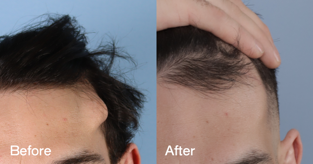
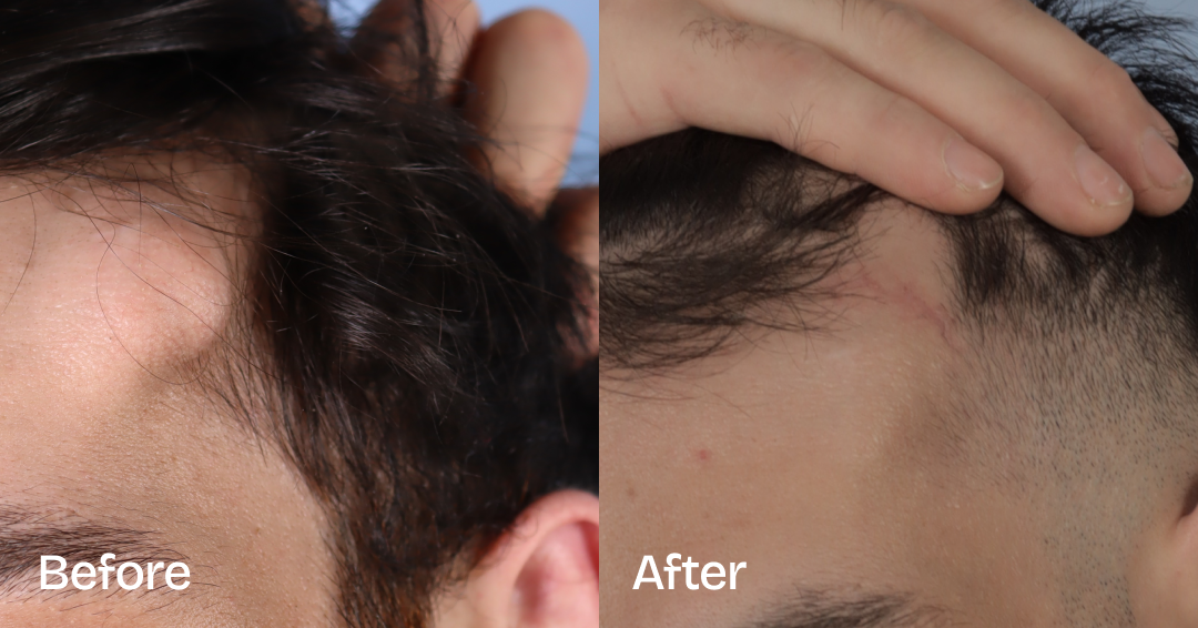
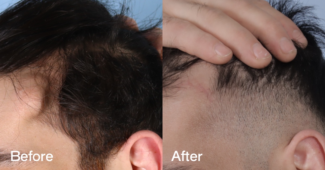
Before and After Photos
The images provided below give examples of real patients who have undergone Osteoma Removal at the CREO Clinic. These results are illustrative of the kinds of results you may expect from the surgery, but outcomes may vary between patients according to individual healing capacity and the type of techniques used.
Is Osteoma Removal Right for You?
Suitability for osteoma removal surgery is assessed during a one-to-one consultation with Dr Tillo. The criteria below offer a general indication only; final eligibility can only be confirmed following clinical assessment.
Who It Is For
– Have a confirmed or suspected osteoma that has been assessed by a specialist, or are seeking specialist assessment of a visible bony prominence
– Are experiencing physical discomfort, pressure, or irritation from the osteoma (usually from glasses frames, helmets, headwear, or contact with surrounding tissue)
– Have a visible prominence that is causing concern and has not been resolved or changed significantly over time
Are in good general physical and psychological health
– Have realistic expectations of what osteoma removal surgery can achieve, including an understanding of scarring and healing timelines
– Are 18 years old or above, or have parental consent where applicable
Who It May Not Be Right For
– Have a recently identified or rapidly growing lump that has not yet been investigated
– Are seeking the removal of an asymptomatic and stable osteoma where the degree of visible prominence is minimal; monitoring may be more appropriate in these cases
– Use nicotine products and are unable to abstain for at least two weeks before and after surgery
– Have underlying health conditions that may increase surgical risk
– Do not have realistic expectations of outcomes, including the permanence of scarring and the possibility of requiring revision
Procedure Variants and Alternatives
The treatment plan for removing an osteoma can vary according to the location of the growth. The three most frequently treated areas at the CREO Clinic are outlined below.
Forehead Osteoma Removal
For forehead osteoma, the endoscopic approach can allow for incision concealment within the hairline, although this is typically suited to smaller osteomas. A larger osteoma may require a direct excision that will also leave a smoother, more even forehead contour, but with a small scar.
Skull Osteoma Removal
A CT scan may be necessary when planning a removal across the scalp.
Osteoma Removal Near the Ear
Depending on the osteoma’s position around the ear, the removal scar may be hidden behind the outer ear itself.
Benefits of Osteoma Removal Surgery
There are multiple key benefits that may follow osteoma removal surgery, depending on the osteoma’s size, location, surgical approach, scarring, and individual healing. Below, we outline some of the more prominent benefits our patients have expressed after the surgery.
Improved Facial or Cranial Contour
The removal of a benign osteoma may improve the contour of the forehead or skull profile. Osteomas can disrupt an otherwise smooth facial contour, and the removal is designed to reduce this bony prominence where clinically appropriate.
Reduced Visible Prominence
A pronounced osteoma can create a visible lump on the skull, which may affect the surface contour of the forehead or scalp. By working to flatten out the osteoma, a removal surgery can help even out the surface of the forehead or skull. The evenness of the surface is often dependent on the size of the osteoma, its location, the surgical approach used, scar formation, and the degree of contour change achievable as a result.
Improved Comfort and Practicality
Direct contact with an osteoma, such as pressure from a hat, helmet, or glasses frames, can sometimes cause localised tenderness or even headaches. Discomfort as a result of osteoma may be relieved through osteoma removal surgery, helping to smooth out the surface and reduce tenderness on the forehead and skull.
Reassurance Following Specialist Assessment
The surgeon-led consultation process at CREO can provide a clear diagnosis of an osteoma, with imaging recommended where clinically appropriate to support assessment and guide next steps. For many patients, an osteoma diagnosis helps to confirm the benign nature of their growth and allows them to move forward with an appropriate treatment.
The CREO APEX Recovery System
What to Expect: Before, During and After
At the CREO Clinic, we work to keep surgical journeys consistent to allow our patients to have a clear picture of what to expect from osteoma removal. The stages below outline the preparation for the surgery, the surgery itself, and the nature of recovery and aftercare needs.
Before Surgery
Stop nicotine consumption: Abstain from smoking, vaping, and all nicotine products for at least two weeks before surgery, as nicotine may impair wound healing
Review your medications: Inform Dr Tillo of all medications and supplements you are taking, including blood thinners and anti-inflammatories. Some may need to be paused before surgery; do not stop any prescribed medication without first consulting your GP
Attend any pre-operative imaging appointments: Where a CT scan or X-ray has been recommended as part of surgical planning, ensure this is completed
Arrange transport and support: Local anaesthetic procedures allow for an earlier return home than general anaesthetic, but you should avoid driving immediately afterwards
Day of Surgery
Initial Recovery
Most patients are able to return to desk work within one to two days following local anaesthetic procedures, and within a few days following general anaesthetic procedures, depending on individual recovery. Activities that risk contact with or pressure on the treated area should be avoided for several weeks. This includes wearing tight-fitting glasses frames, helmets, or headwear that sits directly over the surgical site.
Laser scar management treatments are included as part of the APEX aftercare programme and are commenced as soon as clinically appropriate. A follow-up wound check will be arranged for seven to ten days following surgery.
Final Results
Risks, Limitations and Considerations
As with any surgical procedure, osteoma removal carries recognised risks. All risks will be discussed in detail during your consultation with Dr Tillo, and surgery will only be recommended where the clinical case supports it.
Some potential risks and considerations include:
- Scarring
- Swelling and bruising
- Asymmetry or contour irregularity
- Changes to sensation or temporary numbness in the surrounding skin
- Wound breakdown or delayed healing
- Infection
- Haematoma or seroma formation
- Adverse reaction to anaesthesia
- Incomplete removal, requiring revision
- Recurrence (estimated at less than 1% following complete excision)
Costs and Financing Options
The cost of osteoma removal at the CREO Clinic starts from £3,700. Prices can fluctuate according to the complexity of the surgery, and a final all-inclusive quote can be provided only after a clinical consultation with Dr Tillo.
What Determines the Cost of Osteoma Removal Surgery?
There are multiple contributing factors to the price of osteoma removal surgery. Some key contributors are the size of the osteoma, as a larger growth requires longer surgery and more time in the theatre. Location and complexity also contribute to time in theatre, which is the primary cost driver.
Any imaging performed as a diagnostic tool will also contribute to the final cost of surgery.
Financing Options
The CREO Clinic offers interest-free loans over 12 months via its financing partner, Chrysalis Finance. Longer-term payment plans are available at increased rates of interest. All borrowing is subject to approval.
Why Choose CREO Clinic
The CREO Clinic is structured around clinically-led surgical planning, with specialist assessment and treatment planning tailored to the patient’s anatomy, osteoma characteristics, and suitability for osteoma removal. Our fully integrated aftercare helps ensure that treatment is continuous throughout recovery, working to create consistency and structure during healing and follow-up.
Specialist Anatomical Expertise
At the CREO Clinic, all procedures are performed by specialist plastic surgeons with in-depth knowledge of the procedures they perform. This core principle of the clinic helps support detailed assessment, anatomical planning, and appropriate surgical decision-making across treatments. All our osteoma removals are performed by Dr Omar Tillo, a specialist plastic surgeon who has performed hundreds of osteoma removals over his career.
Consultation-Led Decision Making
All decision making at our clinic is made only after a one-to-one consultation. This ensures that clinical decisions are guided by patient suitability, surgical risk, anatomy, and treatment goals, rather than commercial considerations.
Integrated Aftercare and Recovery Support
Aftercare is fully integrated into every treatment plan at the CREO Clinic, with supplementary treatments such as advanced sutures to optimise healing, our CREO recovery pack including Bio-Oil, Hibiscrub, dressings and immune-supportive vitamins, and laser scar treatment performed as standard through our APEX aftercare system. We believe appropriate recovery steps must be taken from the preparation stage, as they are an important part of wound healing, scar care, and structured recovery support.
Trusted by Patients Seeking Specialist Care
Our patients have consistently given positive reviews across verified review platforms such as RealSelf, where CREO has received an average rating of 4.9 out of 5 stars from contributing patients. We have built trust by providing clear, clinically-led feedback to patients during consultations, and only recommending surgery where it is clinically indicated, and in the interest of achieving patients’ clinical goals.